The advanced characterization of biological samples and innovative materials relies on integrating bio-imaging and nanomechanical technologies.
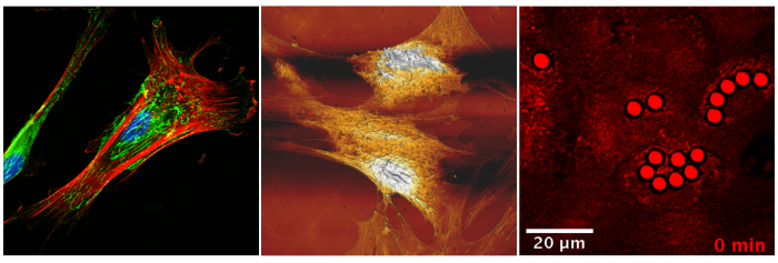
Using a non-linear optical microscope allows the simultaneous combination of two-photon excitation (TPEF), second harmonic generation (SHG), and coherent anti-Stokes Raman scattering (CARS). This approach is ideal for high-resolution dynamic studies on living cells.
Simultaneously, the atomic force microscope for biological applications (BioAFM) enables accurate topographical analysis and the measurement of local mechanical properties. Through Force Spectroscopy, parameters like Young's modulus can be determined directly in a physiological environment.
This synergy of morphological, topographical, and mechanical investigations provides crucial tools for developing cutting-edge solutions in nanomedicine, regenerative medicine, materials science, and in the pharmaceutical industry.